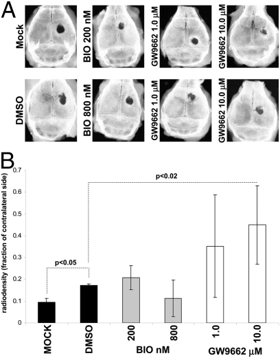
Fig. 6.
Three-millimeter-diameter calvarial defects were induced in nude mice. One million hMSCs pretreated with BIO or GW were mixed with plasma and administered to the bone lesion. Subsequent doses were injected at 14-day intervals until day 50 (Fig. S6C). (A) X-rays of explanted crania. For the GW group, specimens representing the range of the standard deviations are presented. (B) The ratio of lesioned to contralateral (intact side) radio-opacity calculated by image analysis software. Data are means ± SD (n = 6, n = 5 for mock).

