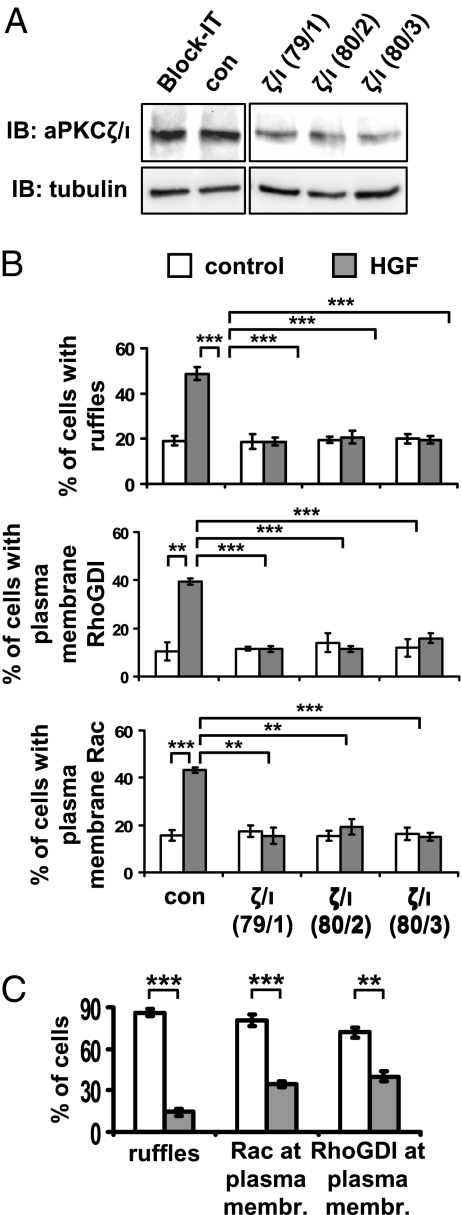
Fig. 4.
aPKCζ/ι mediates both HGF- and myr-DGKα-induced extension of membrane protrusions. (A) MDCK cells were transfected with three combinations of PKCζ (79, 80) and PKCι (1–3) specific siRNAs. Whole-cell lysates were analyzed for levels of aPKCζ/ι expression by western blot. (B) MDCK cells were transfected as in A, treated with 10 ng/mL HGF for 15 min, fixed, and stained for Rac or RhoGDI and actin. C, 110 cells, analyzed for the presence of ruffles and Rac or RhoGDI at the plasma membrane. n = 4 (Rac and RhoGDI), n = 8 (ruffles), with SEM; **P < 0.005, ***P < 0.0005. (C) MDCK cells were transiently transfected either with myr-DGKα alone or cotransfected with myr-DGKα and PKCζKW, cultured overnight in the absence of serum, fixed, and stained for myc and flag tags, actin, and Rac or RhoGDI. C, 20 transfected cells, scored for the presence of ruffles and Rac or RhoGDI at protrusion sites. n = 4, with SEM; **P < 0.001, ***P < 0.0001.

