Fig. 5.
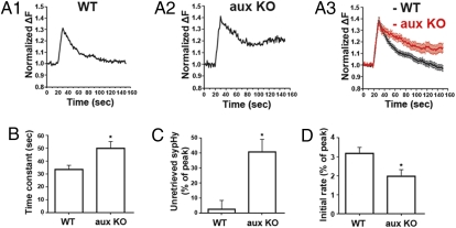
Auxilin knockout reduces the rate of endocytosis. (A) Representative examples of synaptopHluorin signal from a single hippocampal bouton (A1 and A2) and the averaged fluorescence trace (A3); 77 boutons from 4 wild-type mice and 48 boutons from 4 auxilin knockout mice after 200 AP at 20 Hz in WT (black) and auxilin KO (red) neurons. Traces are normalized to the basal fluorescence recorded before stimulation. The decay of the fluorescence signal indicates endocytosis. (B) The endocytic time constant of hippocampal boutons after 200 action potentials at 20 Hz in WT mice (n = 27) and auxilin KO mice (n = 14); *P = 0.01. (C) The percentage of fluorescence that remained unretrieved after 120 s in WT and auxilin KO mice; *P = 0.0045. The data were normalized to the peak of the fluorescence increase. (D) The initial endocytosis rate within 10 s after stimulation in WT and auxilin KO mice; *P = 0.0184. Error bars are SEM.

