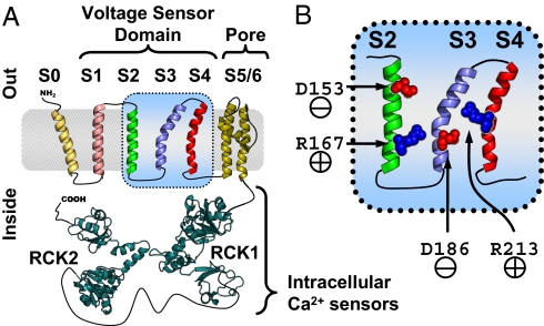
Fig. 1.
BKCa α subunit topology and voltage-sensing residues. (A) BKCa channel α subunit membrane topology (12) and putative structure (intracellular Ca2+ sensing RCK1 and RCK2 by homology to a bacterial RCK domain (19). (B) Close-up of the BKCa voltage sensor domain (VSD) segments S2–S4, indicating the approximate positions of voltage-sensing residues D153 (S2) and D186 (S3) in red and R167 (S2) and R213 (S4) in blue (44).

