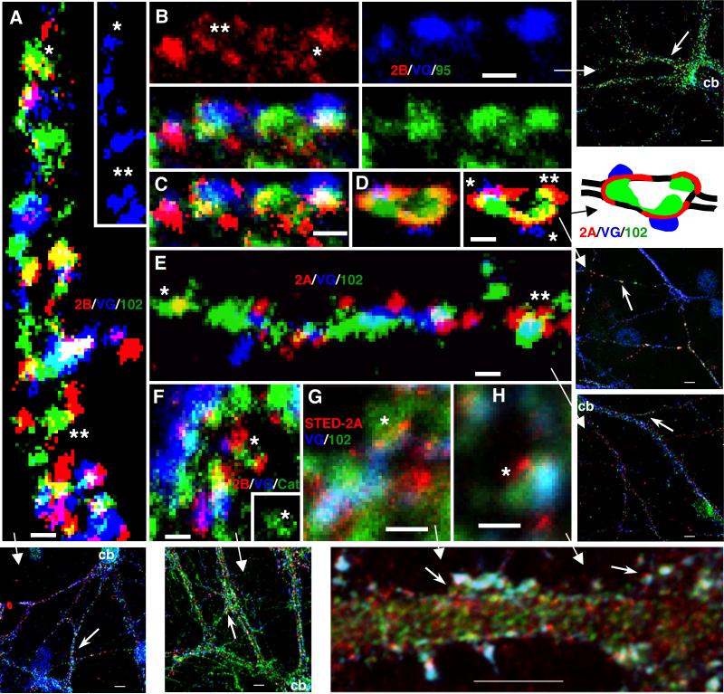
Figure 2.
High magnifications of parts of micrographs (additionally processed for brightness and contrast) from triple immunofluorescence labeling of hippocampal cultures with 1) live surface labeling of NR2A (red; 555 for D,E; 647 [STED] for G,H) or NR2B (red-555; A-C,F), 2) the presynaptic marker VGLUT (blue; 647 for A,D-H; 488 for B,C), and 3) a third protein (green; 488 for A,D-H; 647 for B,C) including PSD-95/93 (B,C), SAP102 (Neuromab; A,D,E,G,H), or catenin (F). A. NR2B/SAP102: note colocalization in areas devoid of VGLUT labeled terminals (shown also alone in the inset)-some appear to be real associations (*) while others may be coincidental (**). B,C. NR2B/PSD-95/93: NR2B forms in a perisynaptic ring (**) around synaptic PSD-95/93 and the terminal, and forms a ring of 3 puncta around an extrasynaptic punctum of PSD-95/93 (*); C is the same image, with the NR2B labeling shown in higher contrast. D. NR2A/SAP102: this is an enlarged region found along a thin, distal dendrite. Note how NR2A labeling is spread in the perisynaptic regions surrounding 2 synapses (*)-SAP102 forms around the enlargement in conjunction with both the synaptic and extrasynaptic (**) NR2A; the right image is a high contrast version of the left one. In the diagram, the outline of the thin dendrite is shown as black lines. E. NR2A/SAP102: NR2A forms a partial perisynaptic ring around synaptic SAP102 (**); in another example (*), the colocalization of NR2A with an elongate punctum of SAP102 may be coincidental (see text). F. NR2B/catenin: 2 NR2B puncta, one synaptic and one extrasynaptic, associate with an elongate punctum of catenin (*; seen alone in the inset). G,H. NR2A[STED]/SAP102: synaptic or perisynaptic puncta (*) of NR2A can be seen at slightly higher resolution than in the confocal images. Scale bars are 500 nm.

