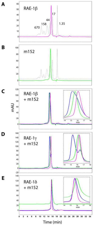
Figure 1. m152 binds to RAE-1 directly.
A,B, Purification of RAE-1 and m152. The ectodomains of RAE-1 isoforms and of m152 were expressed and purified as described in Materials and Methods. Shown here are the chromatographs of RAE-1β (A, pink) and m152 (B, green) overlaid with the protein molecular weight standards (dashed line). C,D,E, Binding of m152 to RAE-1β, γ, and δ examined by a SEC binding assay. m152 and RAE-1 were mixed in equimolar amounts, incubated for 30 min on ice, analyzed chromatographically (blue), and compared to the profile of individual m152 (green) and RAE-1 proteins (pink) alone. Insets show enlargements of the peak regions. (Flow rate was 0.75 ml/min, so elution volumes may be calculated from the indicated time of elution.)

