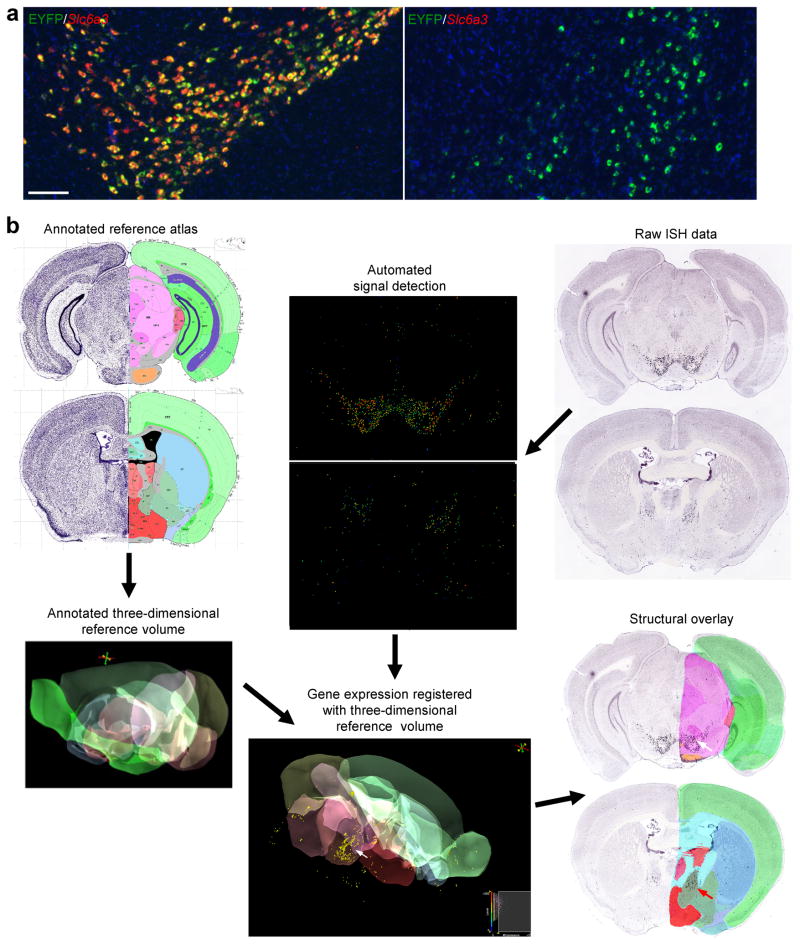
Figure 3.
Informatics processing of the ISH characterization data. (a) Co-labeling of EYFP (green) with Slc6a3 (red) in the Slc6a3-Cre/Ai3 mouse by DFISH. In the VTA/substantia nigra region (left panel), EYFP was largely co-localized with Slc6a3 (>90% cells were co-labeled), demonstrating expected Cre-recombination in dopamine neurons. In the basal forebrain area (right panel), the cluster of EYFP-positive cells were Slc6a3-negative. Scale bar, 200 μm. (b) EYFP ISH data from a Slc6a3-Cre/Ai3 mouse were digitized and registered to the Allen Reference Atlas, which allows anatomical search and comparison. Two sections of registered EYFP ISH data are shown overlaid with anatomical partitioning. EYFP expression is present in both sections (white and red arrows).

