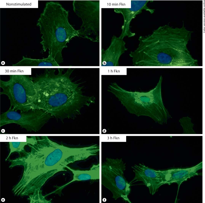
Fig. 1.
a Background staining of F-actin in nonstimulated HUVECs. Cells stimulated with 1 n M Fkn and stained for F-actin after 10 min (b), 30 min (c), 1 h (d), 2 h (e), and 3 h (f) are shown for comparison. Stimulation with 10 n M Fkn resulted in a nearly identical time course of F-actin reorganization (data not shown). Original magnification × 1,000.

