Fig. 3.
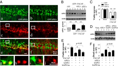
Knockdown of APE1 abrogates PACAP neuroprotection of CA1 following GCI. (A) CA1 was cotransfected for 14 d with lentiviral vectors expressing GFP and the APE1-targeting sequence APE1t (b) or the scrambled sequence APE1s (a), and the sections were immunostained for APE1 (c and d). e (low magnification) and g and h (high magnification) show double-label immunofluorescence of APE1 (red) and GFP (green) in GFP/APE1s-transfected CA1 neurons. (Scale bar, 30 μm.) f (low magnification) and i and j (high magnification) show reduced APE1 expression (red) in GFP/APE1t-transfected CA1 neurons. (B) Western blots show the expression levels of APE1 in CA1, at 14 d after transfection with either GFP alone (Lanes 1–4) or GFP together with APE1t (Lanes 5 and 6) or APE1s (Lanes 7 and 8), without or with PACAP infusions (0.2 nmol, x 3 times, −24, −12, and −6 h). The graph illustrates the relative changes of APE1 expression in CA1 after PACAP treatment. **, P < 0.01; n = 6 per group. (C) Knockdown of APE1 expression attenuated CA1 cell survival after GCI. Viable CA1 neurons were quantified at 4 d after GCI or sham operation after PACAP infusions (0.2 nmol, 24, 12, and 6 h before and 0.5, 6, and 24 h after GCI). **, P < 0.01; n = 8–9 per group. (D) Western blots show the expression of APE1 in CA1, either nontransfected (Lanes 1 and 2) or at 14 d after transfection with APE1t (Lanes 3 and 4), cotransfection with APE1t/hAPE1 (Lanes 5 and 6), or cotransfection with APE1t/hAPE1m (Lanes 7 and 8). (E and F) Overexpression of human APE1, but not the DNA repair-incompetent hAPE1m, restored the prosurvival effect of PACAP for CA1 after GCI. Viable (E) and DNA-damaged CA1 neurons (F) were quantified, respectively, at 4 d after GCI. *, P < 0.05; n = 8–9 per group.

