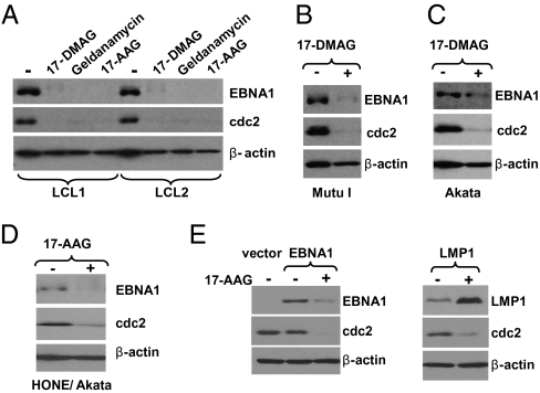
Fig. 1.
Hsp90 inhibitors decrease expression of EBNA1. (A) LCL cells (line 1 and 2) were treated with no drug, 17-DMAG (0.17 μM), geldanamycin (0.5 μM), or 17-AAG (0.5 μM) for 96 h. (B and C) Mutu I and Akata Burkitt lymphoma cells were treated with no drug or 17-DMAG (0.17 μM) for 96 h. (D) The EBV-infected NPC cell line HONE/Akata was treated with no drug or 17-AAG (0.5 μM) for 48 h. (E) AGS gastric cells (EBV-negative) were transfected with empty vector (pSG5), pSG5-EBNA1, or pSG5-LMP1 as indicated, followed by a 48-h treatment with 17-AAG (0.25 μM) beginning at 4 h after transfection. Whole-cell extracts were prepared and immunoblot analysis was performed to analyze the expression of EBNA1, cdc2 (a known cellular substrate of Hsp90), cellular β-actin, or LMP1 as indicated.

