Fig. 3.
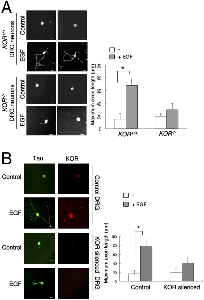
KOR is required for EGF-induced axonal extension of primary DRG neurons. (A) Two immunohistochemistry images of KOR+/+ and KOR−/− primary mouse DRG neurons treated with, or without, EGF for 48 h and detected with anti-Tau antibody. Quantitative analysis of axon length by scoring 50 neurons is shown on the right (*P < 0.05). (Scale bars: 25 μm.) (B) Immunohistochemistry of rat DRG neurons transfected with control siRNA (first and second panels from the top) or KOR-specific siRNA (third and fourth panels from the top) on control (first and third panels from the top) or EGF treatment (second and fourth panels from the top), stained with anti-Tau or anti-KOR antibodies. Quantitative measurement of axon length from 50 neurons is shown on the right (*, P < 0.05). (Scale bars: 25 μm.)

