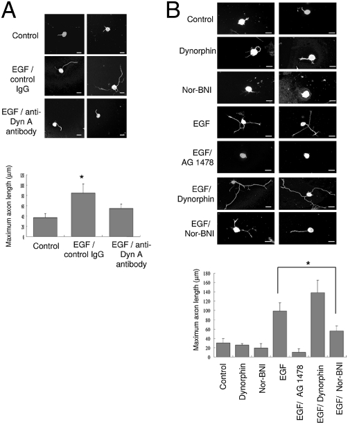
Fig. 4.
KOR ligand is required for EGF-induced axon extension in primary DRG neurons. (A) Two immunohistochemical images from primary rat DRG neurons treated with EGF and anti-dynorphin A antibody (Bottom). Untreated neurons (Top) or neurons treated with IgG and EGF (Middle) serve as controls. Quantitative analysis of axon length from 50 neurons of each experiment is shown on the bottom (*, P < 0.05). (B) Two immunohistochemical images from primary rat DRG neurons treated with dynorphin, nor-BNI, EGF, EGF plus AG 1478, EGF plus dynorphin, and EGF plus nor-BNI. Quantitative analysis of axon length from 50 neurons of each experiment is shown below (*, P < 0.05). (Scale bars: 25 μm.)

