Fig. 1.
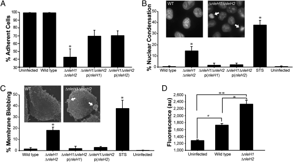
Cells infected with EPEC ΔnleH undergo apoptosis. Quantification of live adherent HeLa cells (A), nuclear condensation (B), membrane blebbing (C), and cytoplasmic Ca2+ (D) following infection of HeLa cells with WT EPEC, ΔnleH1ΔnleH2 mutant, and p(nleH1) or p(nleH2) complemented strains. The number of adherent cells was significantly reduced in cells infected with EPEC ΔnleH1ΔnleH2 (*) compared with uninfected cells and cells infected with WT EPEC. Complementing the mutant with either p(nleH1) or p(nleH2) partially restored the WT phenotype (A). The number of cells displaying nuclear condensation or fragmentation (B) or membrane blebbing (C) was significantly higher in HeLa cells infected with ΔnleH1ΔnleH2 or after treatment with STS as a control (*) compared with cells infected with EPEC WT or the complemented mutant strains. Although significant Ca2+ release was observed in WT EPEC-infected cells compared with uninfected cells (#), HeLa cells infected with the ΔnleH1ΔnleH2 had significantly elevated cytosolic Ca2+ levels compared with cells infected with WT EPEC (*) or uninfected cells (**) (D). In A, B, and C, the nonparametric Mann-Whitney test was used to determine significance, because the data did not follow Gaussian distribution. *P < 0.05. In D, significance was tested using the one-way ANOVA Bonferroni multiple-comparisons test. *,#,**P < 0.05.

