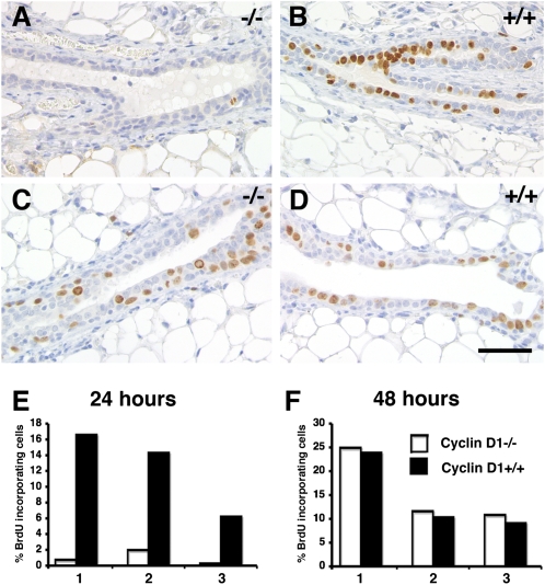
Fig. 2.
Cyclin D1- and progesterone-induced proliferation. (A and D) Mice engrafted with cyclin D1−/− and WT epithelia were stimulated with progesterone for 24 h (A, B, and E) or 48 h (C, D, and F). Histological sections of contralateral mammary glands engrafted with cyclin D1−/− (A and C) or WT (B and D) epithelia and stained with an anti-BrdU antibody. (Scale bar: 40 μm.) (E and F) Bar graphs showing BrdU incorporation in cyclin D1−/− and WT MECs ± SEM (n = 3), 1,000 cells counted per mouse.

