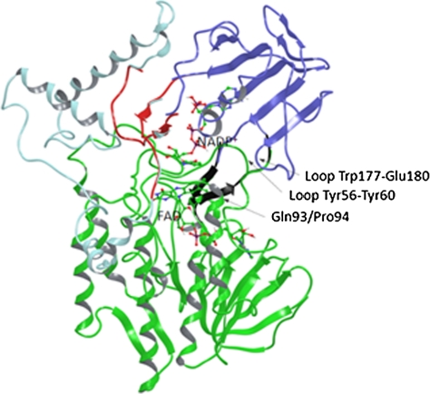
Fig. 1.
Cartoon representations of the different domains in the PAMO structure. FAD domain is shown in Green, Dark Blue corresponds to the NADP-binding domain, and Light Blue to the helical domain. The NADP- and FAD-binding domains are linked by two antiparallel β-strands in a hinge-like structure configuration (Black). NADP-binding and helical domains can be considered as separate entities connected by two segments (Red). The randomization site Gln93/Pro94 in the N-terminal region of the α-helix is also labeled.

