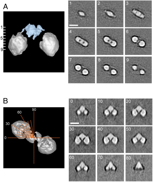
Fig. 4.
Sections through Keap1 molecule. (A) Horizontal slices perpendicular to the pseudo twofold symmetric axis show the characteristic cherry-shaped structure of Keap1 (Right). Positions of the cross sections, at 9.43 Å intervals throughout the molecule, are numbered from one to nine on the side surface view (Left). The stem structure, occupying 13.5% of the total volume (Blue). (B) Axial slices (Orange Dotted Lines; top surface view) every 10 ° from 0 ° to 90 ° (Left). Each sectional image is shown with corresponding angle (Right). Two globular domains are connected to the central stem-like density with slender linkers. The central stem-like density comprises two thin layers connected to one another but separated by a thin gap. The globular domains on either side are dense cylinders with round corners, pierced by an apparent low-density tunnel between the two surface orifices. Protein is displayed in bright shades. Scale bars, 100 Å.

