
Fig. 3.

Formation of engineered vessels in vivo. (A–D) Networks formed by HUVECs imaged with confocal laser-scanning microscopy at 4 weeks (A) without hMSCs, (B) with standard condition hMSCs, (C) with hMSCs cultured with TGF-β, and (D) with hMSCs cultured in low serum MesenPro medium. For (D) only, maximum intensity projection of 30 consecutive frames (1 sec) was used to demonstrate flow of DiD-labeled blood cells. H&E and human-specific CD31 immunohistochemical staining of scaffolds explanted 4 (E–L) and 11 (M–T) days after implantation. Acellular regions (*) derive from scaffold. EC-derived tubular structures have formed by 4 days (arrowheads) but do not contain blood cells indicating that they are not yet functional. By 11 days, the structures were larger with more robust CD31 staining, and most lumens were filled with blood cells (arrows). The symbols mark only a representative sample of indicated structures. (Scale bars: 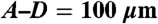 ;
; 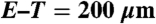 .)
.)