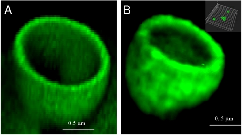
Fig. 1.
Confocal microscopy images of 1 μm fluorescently labeled capsules with shells comprising of PLL and PGA without embedded proteins (A) or after incorporation of BMP2 and TGFβ1 and dissolution of the core: PLL-(PGA-PLL)2-BMP2-PLL-TGFβ1-PLLFITC (fluorescein isothiocyanate) (B). The hollow cross-section of the capsules is depicted. The Inset shows dispersed capsules.

