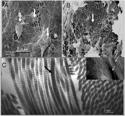
Fig. 3.
SEM images of osetoblasts from EBs growing in the presence of PEI-(PGA-PLL)2-BMP2-PLL-TGFβ1-PLL particles. (A): Osteoblast visualizations; (B): mineralized collagen fibers (zoom × 40). In the presence of the multilayered particles without BMP2 and TGFβ1, osteoblasts or bone matrix were not observed. (C) TEM observation after in vivo bone induction by EBs growing in the presence of the alginate gel and PLL-(PGA-PLL)2-BMP2-PLL-TGFβ1-PLL particles: bundle of collagen fibrils seen in longitudinal and transverse section. The arrows indicate the type I collagen fibrils viewed in longitudinal section. Note the characteristic cross striation and periodicity of this type of collagen (67 nm/0.5 μm). The mineralization of the collagen fibers is also visualized in the Inset.

