Fig. 3.
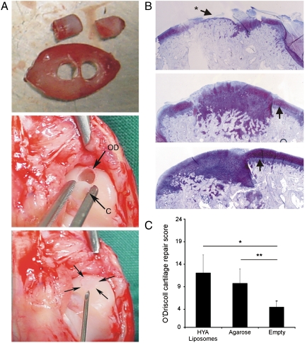
(A) Upper: to show the size of IVB cartilage, an example is shown in which two cartilage grafts 3 mm in diameter each were cored out. For transplantation purposes onl,y one graft was cored out of the center of the IVB cartilage. Middle: an IVB cartilage graft (C) is press-fit implanted in an osteochondral defect (OD) of medial femur condyle. Lower panel: Anatomy of a repaired osteochondral defect directly after implantation of an IVB cartilage graft (indicated by Arrows). (B) Repair of osteochondral defects. Thionine staining of medial condyle sections 9 mo after creation of an osteochondral defect. Upper: top-view of an empty, non-grafted defect. Note the penetration of hypertrophic subchondral bone in the empty defect and into surrounding cartilage layers indicated by the Arrow marked with an Asterisk. Middle and Lower: Representative examples of defects transplanted with IVB cartilage from IVB filled with HA-TGF-β1/Suramin gel (Middle) and agarose (Lower). Incompletely differentiated mesenchyme is integrated laterally and a superficial layer with more intense thionine staining can be distinguished (Arrows). The Arrows indicate the original joint cartilage. Penetration of subchondral bone into the joint of the implanted IVB graft was never observed. Magnification: 25X. (C) O’Driscoll scores for cartilage repair. After implantation with a cartilage graft from the IVB using agarose or HA-TGF-β1/Suramin gel, osteochondral repair scores (9.8 and 12.1, resp.) was significantly (∗∗ p = 0.003 and ∗ p = 0.001, resp.) better compared to empty defect (average repair score 4.5).

