Fig. 3.
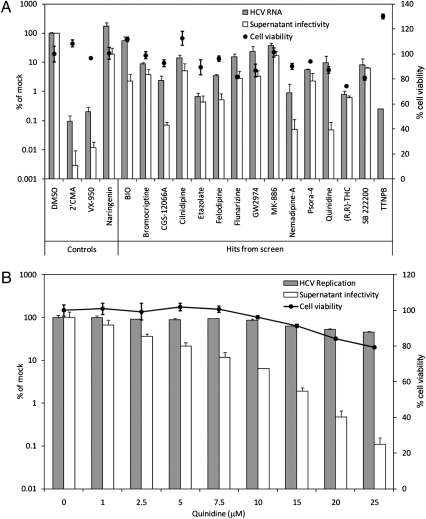
Leading inhibitors of HCV replication and infectious virus production from small-molecule library screen. (A) Intracellular HCV RNA levels and supernatant infectivity 96 h after electroporation of Huh-7.5 cells with the genomic RNA of Jc1 HCV and treatment with 15 leading replication/infectious virus production inhibitor hits at the concentrations (SDC) indicated in Table 1. Intracellular HCV RNA levels and supernatant infectivity were determined by quantitative RT–PCR and a cell-viability–adjusted median tissue culture infectious dose (TCID50)/mL assay, respectively. Note that a value is not shown for the supernatant infectivity of TTNPB-treated cells because no cells were infected at any of the serial dilutions of this supernatant. For inhibitor cytotoxicity determination, cell viabilities of Huh-7.5 cells electroporated with carrier (water) before inhibitor treatment were measured by CellTiter-Glo assay. Each point is the mean ± SD of at least three independent experiments. (B) Quinidine dose-dependently inhibits infectious HCV production in Huh-7.5 cells. Virus replication and supernatant infectivity were determined by a Gluc reporter HCV-based assay. Cytotoxicity to Huh-7.5 cells electroporated with only carrier was determined as in A. Each point is the mean ± SD of two independent experiments carried out in duplicate. Values in A and B are expressed as a percentage of 0.1% DMSO-treated cells.

