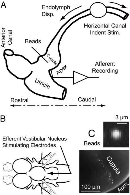
Fig. 1.
Experimental set-up. (A) Schematic of surgically exposed region of the membranous labyrinth showing location of indentation stimulus, single-unit afferent recording, and fluorescent microbead placement. (B) Bipolar stimulating electrodes placed in the brainstem efferent vestibular nucleus. (C) (Lower) Fluorescent microbeads adhered to the cupula. (Upper) Intensity of a single bead viewed through the transparent membranous labyrinth in the living animal.

