
Figure 1.
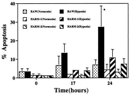
Hypoxia-induced apoptosis in macrophages. RAW and HARM cells grown in RPMI 1640 medium containing 1% FBS were exposed to hypoxia for 24 hr. Macrophage apoptosis was evaluated by fluorescence-activated cell sorting analysis after PI staining. The graph represents the results of five separate experiments. The results are expressed as mean ± SD. ∗, Data sets are different at the 95% confidence level (P < 0.05) compared with normoxic condition.