
Figure 4.
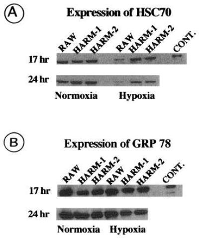
Western blot analysis of heat shock proteins in RAW and HARM cells. RAW and HARM cells exposed to either hypoxia (2% O2) or normoxia (21% O2) for 17 and 24 hr were lysed on ice and extract protein was subjected to electrophoresis in SDS/12% polyacrylamide gels. Gels were electroblotted to nitrocellulose membranes, which were immunoblotted with anti-HSC 70 (A) or anti-GRP 78 (B) antibody conjugated to horseradish peroxidase. Antibody–heat shock protein complexes were detected by chemiluminescence. Bovine brain HSC 70 and recombinant hamster GRP 78 was used as positive controls for HSC 70 and GRP 78, respectively. The figure is a fluorogram.