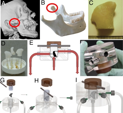
Fig. 1.
Tissue engineering of anatomically shaped bone grafts. (A–C) Scaffold preparation. (A and B) Clinical CT images were used to obtain high-resolution digital data for the reconstruction of exact geometry of human TMJ condyles. (C) These data were incorporated into MasterCAM software to machine TMJ-shaped scaffolds from fully decellularized trabecular bone. (D) A photograph illustrating the complex geometry of the final scaffolds that appear markedly different in each projection. (E) The scaffolds were seeded in stirred suspension of hMSCs, to 3 million cells per scaffold (≈1-cm3 volume) and precultured statically for 1 week to allow cell attachment, and then the perfusion was applied for an additional 4 weeks. (F) A photograph of perfusion bioreactor used to cultivate anatomically shaped grafts in vitro. (G–I) Key steps in bioreactor assembly (see Movie S1).

