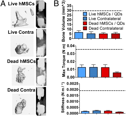
Fig. 5.
Structure/function results from in vivo delivery of live or devitalized QD-loaded hMSCs. (A) Micro-CT (Left) and x-ray (Right) images of the best bone formation per group in defects receiving live hMSCs, acellular scaffold contralateral to live hMSCs, devitalized hMSCs, or acellular scaffold contralateral to devitalized hMSCs. (B) Twelve-week in vivo bone volume and postmortem defect maximum torque and torsional stiffness. Black dashed lines indicate values for the QD-free hMSC treated defects from the segmental defect study without QDs.

