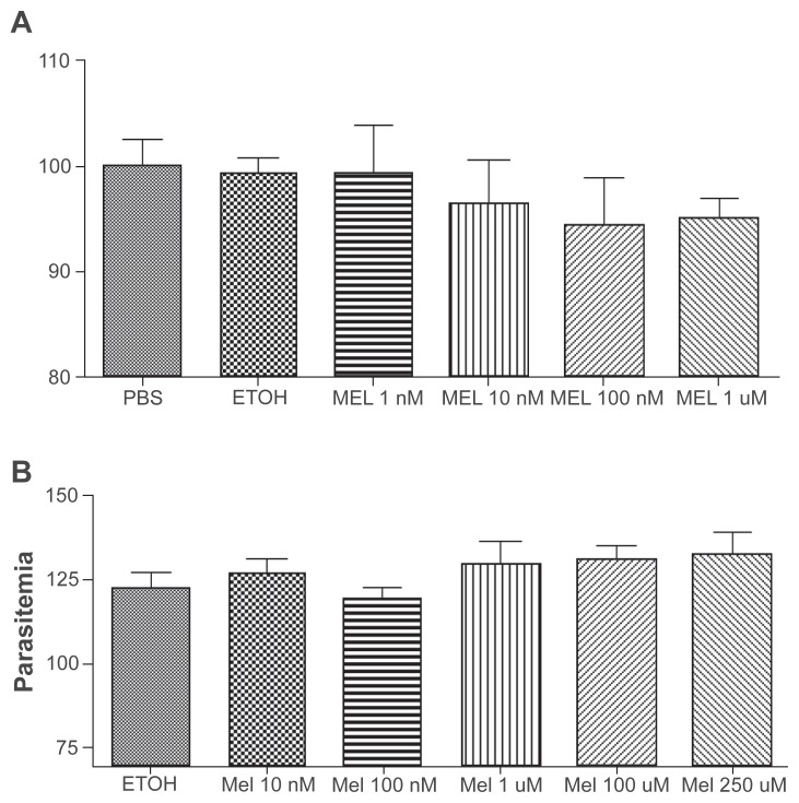
Figure 4.
In vitro culture of P. berghei A) and P. yoelii B) incubated with different melatonin concentrations. The figure shows P. berghei- and P. yoelii-infected red blood cells (iRBC) after 18 or 13 hours incubation, respectively, melatonin concentrations (1 nM, 10 nM, 100 nM and 1 μM). There are no statistical differences in the number of iRBC. The results are presented as the mean of three independent experiments.

