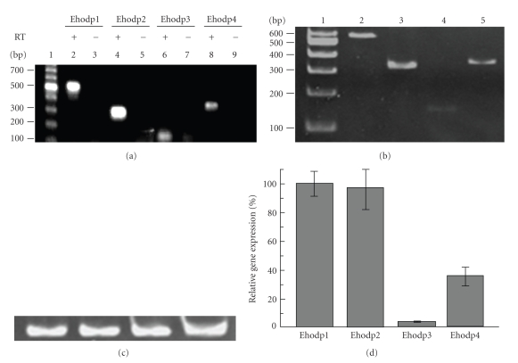
Figure 4.
Expression analysis of the Ehodp1, Ehodp2, Ehodp3 and Ehodp4 genes. (a) RT-PCR products separated by 6% PAGE and ethidium bromide stained. Lane 1, DNA molecular markers (100 bp ladder); lanes 2, 4, 6, 8, amplified 524, 317, 149, and 376 bp DNA fragments (+) corresponding to Ehodp1, Ehodp2, Ehodp3 and Ehodp4 genes, respectively; lanes 3, 5, 7, 9, are the corresponding negative controls (−) for each gene in which no RT was included. (b) Semiquantitative RT-PCR. Gene products were analyzed by 6% PAGE and gels were ethidium bromide stained. Lane 1, size DNA molecular markers; lane 2, amplified 524 bp DNA fragment from Ehodp1; lane 3, amplified 317 bp DNA fragment from Ehodp2; lane 4, amplified 149 bp DNA fragment from Ehodp3; lane 5, amplified 376 bp DNA fragment from Ehodp4. (c) RT-PCR amplification product (222 bp) for actin gene used as control. (d) Densitometry analysis of the Ehodp gene expression shown in (b). Expression of Ehodp1 measured in pixels was considered as 100%. Data is the average of three experiments.

