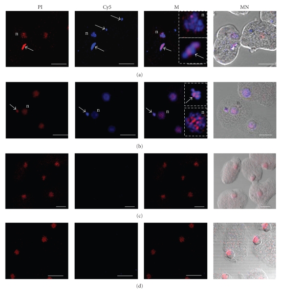
Figure 5.
Ehodp1 gene localization in nuclei and cytoplasmic DNA-containing structures by in situ PCR. Trophozoites of E. histolytica clone A were fixed, permeabilized and used to amplify a specific DNA fragment of the Ehodp1 gene by IS-PCR using Cy5-dCTP. Then, cells were RNase-treated and stained with PI and observed through a laser confocal microscope. (a)–(b) Amplification of Ehodp1 by IS-PCR. (c) Negative control of IS-PCR carried out without Taq DNA polymerase. (d) Negative control of IS-PCR performed without Ehodp1 specific oligonucleotides. (PI) Cells stained with propidium iodide (red channel). (Cy5) Ehodp1 amplification products labeled with Cy5-dCTP (blue channel). (M) Merging of red and blue fluorescent signals. Squares show an image amplification of a nucleus and a cytoplasmic DNA-containing structure. (MN) Merging of fluorescent signals superimposed on the corresponding cellular images obtained by Nomarsky microscopy. Nucleus (n). Arrows indicate cytoplasmic DNA-containing structures. Bar scale corresponds to 8 μm

