
Figure 1.
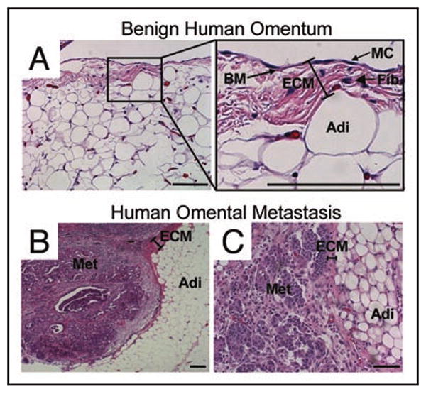
Histology of omental metastasis of human ovarian cancer. Hematoxylin and eosin stain of (A) normal human omentum (MC, mesothelial cells, BM, basement membrane, Fib, fibroblasts, Adi, adipocytes) and (B and C), ovarian cancer omental metastasis (Met, ovarian cancer metastasis). Experimental details were as described in ref.9 Bar in bottom right of each panel = 100 μm in length.