
Figure 2.
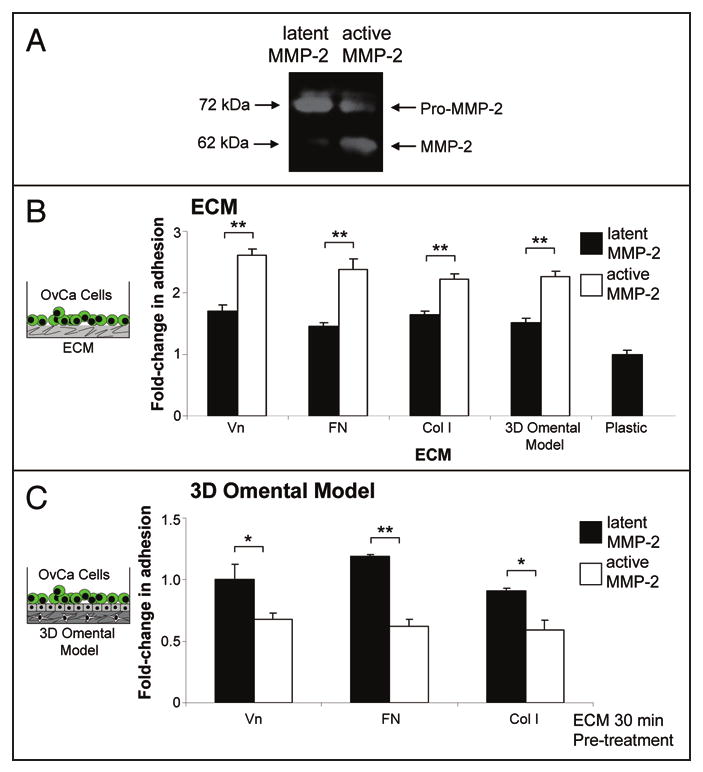
MMP-2 cleavage of vitronectin, fibronectin, collagen I increases primary ovarian cancer cell adhesion. (A) Zymogram. MMP-2 was activated by APMA for 1 hour and was subjected to gelatin zymography.12 (B) Adhesion assay. Full-length Vn (vitronectin), FN (fibronectin) and Col I (collagen I) or the 3D omental model were plated in a 96-well dish. MMP-2 or MMP-2 that was proteolytically activated by APMA was added to the indicated ECM or the 3D model (Cleavage of ECMs was confirmed by silver staining—not shown). After an 1 hour incubation, the wells were washed with serum-free media and fluorescently-labeled primary human ovarian cancer cells were added for 30 min. The wells were washed, fixed and fluorescence measured using a fluorescent plate reader. (B) Competition assay. Primary human ovarian cancer cells were pre-incubated with MMP-2/ECM or APMA-activated MMP-2/ECM for 30 min. in serum-free media. Then the cells were added to the 3D omental model and adhesion quantified as described in (A). *p-value <0.01, **p-value <0.001.