
Figure 3.
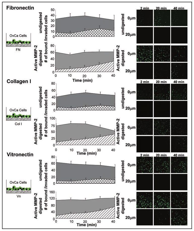
Activated-MMP-2 increases early ovarian cancer invasion through fibronectin, collagen I and vitronectin. Live in vitro imaging of SKOV3ip1 cell adhesion and invasion into fibronectin, collagen I and vitronectin. 200 μg of ECM was plated in a 35 mm glass bottom dish, APMA or APMA-activated MMP-2 added for 1 hour, and the well washed with serum-free media. Fluorescently-labeled SKOV3ip1 cells were added and real-time confocal microscopy images taken at 0 μm depth which represents the surface of ECM and a 20 μm depth below the ECM surface. Representative images at 2, 20 and 40 min. are shown.