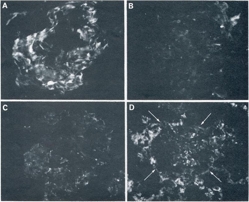
Fig. 3.
The patterns of renal fibrin deposition detected by fluorescein conjugated rabbit antidog fibrin. (A) A glomerulus from sensitized dog 3 studied 10 min following vascularization of the second homograft. Moderate amounts of fibrin are present in an irregular pattern along the glomerular capillary walls (original magnification, × 400). (B) A glomerulus from the same kidney as in A, 60 min after vascularization. Most of the fibrin which was seen at 10 min has disappeared (original magnification, × 400). (C) A glomerulus (10 min) is shown from a homograft placed in nonsensitized control dog. A small amount of fine irregular fibrin is deposited along the glomerular capillary walls. This was the most extensive fibrin deposition seen in any of the control homografts (original magnification, × 400). (D) A glomerulus (arrows) and the surrounding renal tissue are shown from the first homograft (24 hr), placed in a sensitized dog. Moderate glomerular and heavy peritubular fibrin deposits are evident (original magnification, × 250).

