
Fig. 1.
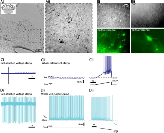
Visualization and spontaneous activity of GlyT2+ neurons in deep cerebellar nuclei (DCN). A Demonstration of GFP in the DCN from GlyT2-eGFP mouse using GFP immunohistochemistry in a paraformaldehyde-fixed cerebellar coronal section. The approximate location of field of view in Ai is marked as a square in the upper left inset; the field of view in Aii is marked as a dashed line square in Ai. Scale bars Ai 100 µm, Aii 20 µm. Arrow in Aii points to a large GFP+ cell; arrowheads point to smaller GFP+ cells. B Typical examples of fluorescence-assisted selection of cells for patch-clamp recordings. Images from exactly the same location in acute slices seen in differential interference contrast (DIC, upper panels) and whole-field epifluorescence (lower panels) configuration. In Bi, several small, GFP-expressing (black asterisk) GlyT2+ cells as well as a non-expressing one (white asterisk) are visible. In Bii, one small and one large GFP+ cell are seen (black asterisks); note that because the large cell in Bii is deeper than the cells in Bi, it is not as clearly visible in DIC even though easily distinguished in fluorescence imaging. C Example recording from an inherently silent GlyT2+ cell. Ci In cell-attached mode only occasional action potentials are seen. Cii After breaking into whole-cell configuration with no bias current (I cmd = 0 pA), the membrane potential of the cell remains below spike threshold until depolarized with current injection (Ciii). In Cii and Ciii, the bottom panel depicts the bias (command) current (I cmd); dashed line in the upper panel marks −50 mV. Traces in Ci–Ciii are from the same cell. Note the different time scale in Ciii. D Example recording from spontaneously active GlyT2+ cells. Di In cell-attached mode, the cell fires spontaneously. Dii After breaking into whole-cell mode with no bias current (I cmd = 0 pA), spontaneous firing continues. Diii When a small amount of hyperpolarizing current (in this example, −10 pA) is injected into the cell, the firing frequency slows down and ultimately ceases. Traces in Di and Dii are from a single cell, Diii is from a different one. Note the different time scale in Diii