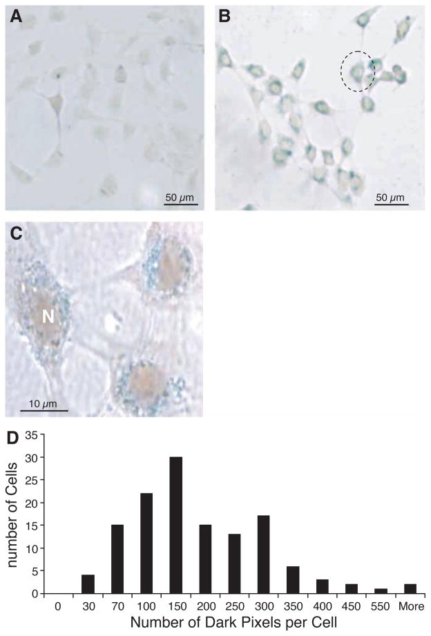
Figure 1.
Optical microscope view stained with Prussian blue to demonstrate the uptake of SPIO particles. (A) No stainable iron was detected in the unlabeled cells. SPIO particles are visible within all cells in (B) as blue iron stain. All of the iron is around the nucleus (N); no intranuclear or extracellular SPIO could be detected (C). (D) Quantification of SPIO particle distribution inside each cell by light microscopy. Note individual cells take up different amounts of SPIO particles (n = 339).

