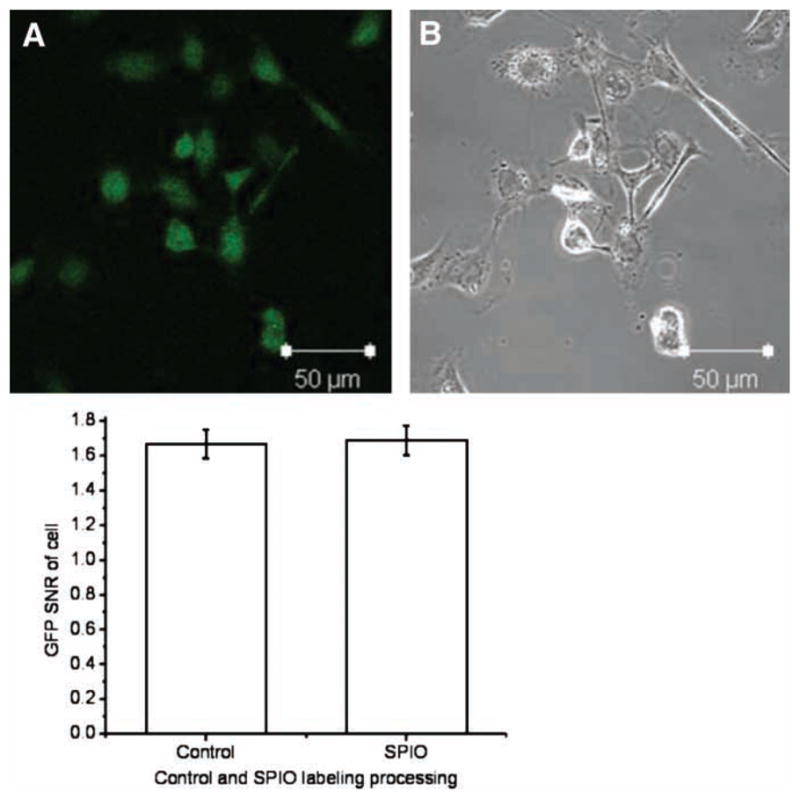
Figure 2.

Confocal microscopic view of labeled cells with Prussian blue staining. (A) GFP expression of whole labeled cells; (B) a light microscope image of the same view demonstrating the uptake of SPIO particles (dark particles in cytoplasm). (C) Quantitative SNR analysis with MetaMorph™ software; no difference was observed in GFP expression between labeled and unlabeled groups, P > 0.05.
