
Figure 1. In vivo excision of Dicer by Col1a1-Cre impairs bone formation at E14.5 and induces embryonic lethality.
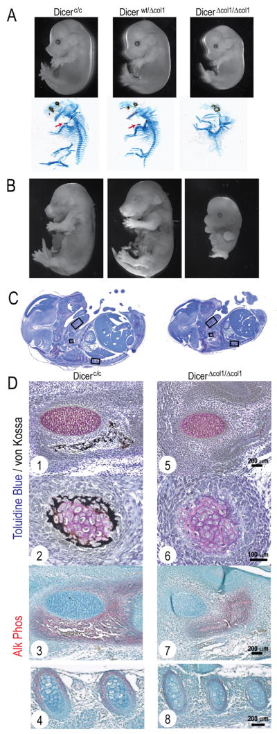
(A) Skeletal deformities observed at E14.5. Upper panel shows smaller but viable DicerΔcol1/Δcol1 embryos at E14.5. Alcian Blue/Alizarin Red staining (lower panel) reveals lack of mineralizing clavicles (see arrow in Dicerc/c and Dicerwt/Δcol1) and a deformed cartilaginous skeleton in DicerΔcol1/Δcol1. (B) Images of E15.5 fetal pups show partial resorption of DicerΔcol1/Δcol1 pup. (C) Embryo sections are compared at E14.5 and (D) detailed (boxed areas) at higher magnification for Dicerc/c (1–4) and DicerΔcol1/Δcol1 (5–8), showing severely impaired mineralization of mandible mesenchyme that surrounds Meckel’s cartilage (1, 5), clavicle (2, 6) and reduced Alk Phos staining in the lower jaw (3, 7) and ribs (4, 8) of DicerΔcol1/Δcol1 embryos.