


Figure 2. Contributing mechanisms for defective osteoblast differentiation in the absence of Dicer.
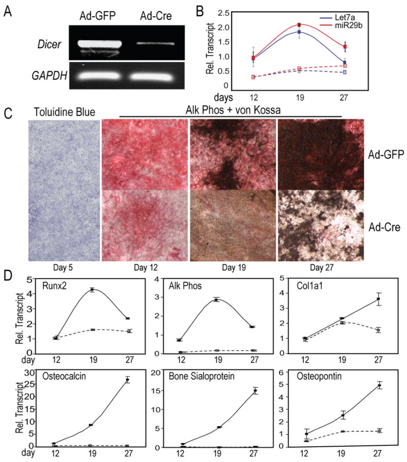
Dicer excision in BMSCs from Dicerc/c mice by Ad-Cre, followed by differentiation into osteoblasts. Ad-GFP was used as control for infection of cells. (A) Genomic PCR of Ad-GFP (control) and Ad-Cre treated cells identified >90% excision of Dicer locus on day 5 (GAPDH, internal control). (B) Lack of miR-29b (Red lines) and let-7a (Blue lines) expression in the absence of Dicer. Solid lines (––) show control Ad-GFP treated cells; dotted lines (---), Ad-Cre Dicer-ablated cells. (C) Cellularity (Tol Blue) was not affected by Dicer excision, but differentiation (Alk Phos activity) and mineral (von Kossa stain) were severely impaired. (D) Expression patterns of early markers of committed osteoprogenitors, Runx2 and Col1a1, and components for the onset and regulation of mineralization by the non-collagenous proteins, OC, BSP and OP, reflect impaired maturation of osteoblasts in Dicer-excised (Ad-Cre) cells.