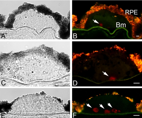
Figure 1.
Immunolocalization of nonfibrillar amyloid oligomers in drusen by the rabbit M204 monoclonal antibody. Sections containing drusen from three individuals are shown. One-micrometer-thick optical images were acquired on an inverted laser scanning confocal microscope. (A, C, E) Differential interference contrast images; (B, D, F) confocal fluorescence images of amyloid oligomer cores (red, Cy3 channel). Arrows: M204-positive cores. Lipofuscin autofluorescence in RPE appeared as an orange-yellow fluorescence. RPE, retinal pigmented epithelium; Bm, Bruch membrane. Scale bar, 10 μm.

