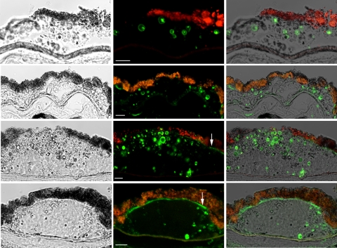
Figure 3.
Mature amyloid fibrils recognized by the WO antibody were localized to the shell of vesicular structures. Shown are representative sections containing drusen from four individuals. Left: differential interference contrast images; middle: WO reactivity (green) reflecting the presence of mature amyloid fibrils; right: overlay of the differential interference contrast and fluorescent images. WO reactivity was seen largely at the surface of vesicular structures. Not all vesicles were stained. Sub-RPE deposit appeared to stain strongly for WO (arrows, bottom). Red-orange granules were due to RPE lipofuscin autofluorescence. Scale bar, 10 μm.

