Figure 1.

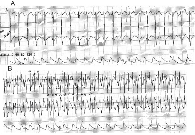
(A) Surface electrocardiogram of a patient in atrial flutter with 2:1 AV conduction. The P waves are not clearly seen, (B) Atrial electrocardiogram of the same patient shows the P waves with 2:1 AV conduction more clearly

(A) Surface electrocardiogram of a patient in atrial flutter with 2:1 AV conduction. The P waves are not clearly seen, (B) Atrial electrocardiogram of the same patient shows the P waves with 2:1 AV conduction more clearly