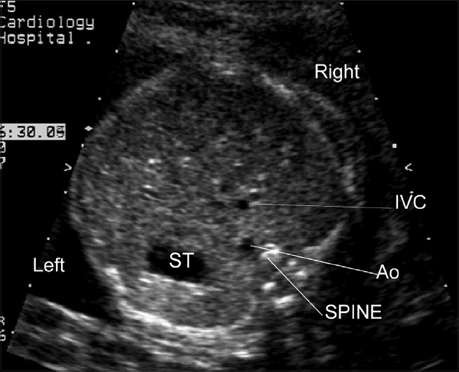
Figure 1(a).

Normal cardiac situs; The fetal stomach (ST) is seen on the left. The descending aorta (Ao) is anterior and to the left of the fetal spine. The inferior vena cava is anterior and to the right of the aorta. (These figures are sequential views commencing with views of the cardiac situs (inferior) and ending with the “three vessel view” in the superior mediastinum)
