Figure 5.
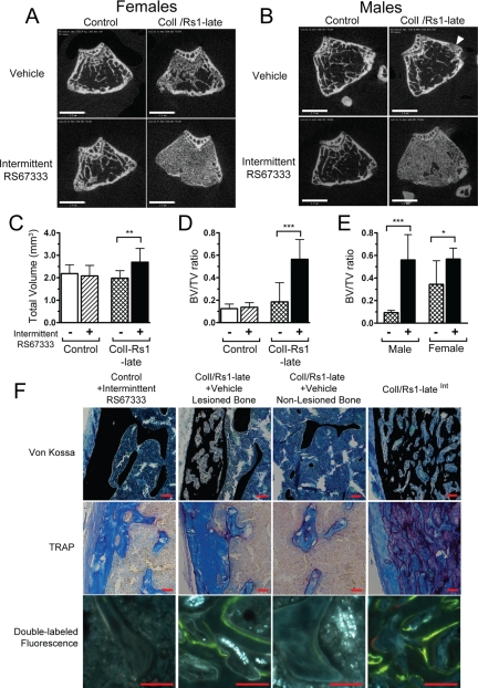
Intermittent ligand activation of Rs1 leads to a localized increase in trabecular bone formation. Representative cross-sectional microCT images through the metaphysis of the distal femur show increased trabecular bone in both (A) female and (B) male ColI/Rs1–late mice, after intermittent RS67333 administration. Note that in the absence of ligand administration the female ColI/Rs1–late mouse shows a small increase in trabecular bone and the male ColI/Rs1–late mouse has a trabecularized region of metaphyseal cortex (arrowhead). Scale bars, 1 mm. C and D, Quantitative microCT assessment of trabecular bone at the distal femur for TV and fractional BV (BV/TV). n = 9 controls + vehicle; 11 controls + RS67333; 11 ColI/Rs1–late + vehicle; 14 ColI/Rs1–lateInt mice. E, Subgroup analysis confirmed that only female ColI/Rs1–late mice showed increased BV/TV in the absence of ligand administration. n = 7 male ColI/Rs1–late + vehicle; 8 male ColI/Rs1–lateInt; 4 female ColI/Rs1–late + vehicle; 6 female ColI/Rs1–lateInt mice. Error bars, mean ± 1 sd. *, P < 0.05; **, P < 0.01; ***, P < 0.001. F, Representative histology of von Kossa, TRAP staining, and fluorescence labeling of longitudinal sections of the femur. Bone from a control animal treated with RS67333 is compared with lesioned and nonlesioned regions of bone from a vehicle treated ColI/Rs1–late mouse and bone from a ColI/Rs1–lateInt mouse. All images shown are from the femurs of male mice, although the histology is consistent with that seen in both sexes. Scale bars, 50 μm.

