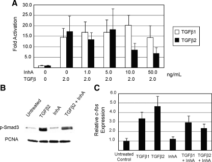
Figure 3.
Inhibin functionally antagonizes TGFβ2 signaling in vitro. A, Luminescent assays for 3TP-lux reporter activation were performed in Y1 adrenocortical cells treated with a fixed amount of TGFβ1 or TGFβ2 (2 ng/ml) and increasing doses of recombinant inhibin-A peptide as indicated. 3TP-lux activity was normalized to the luminescent signal from a constitutively expressed pCMV-Renilla reporter vector that was cotransfected with 3TP-lux into Y1 cells. B, Y1 cells were treated with vehicle (PBS), TGFβ2 (0.5 ng/ml), inhibin-A (100 ng/ml), or TGFβ2 plus inhibin-A for 16 h. Immunoblot analysis was performed on 10–20 μg of protein lysates loaded in equal amounts for each sample. Blots were probed with antibodies to phospho-Smad3 to indicate TGFβ2 signaling activity and proliferating cell nuclear antigen to indicate equal protein loading. C, Y1 cells were treated with vehicle (PBS), TGFβ1 (0.5 ng/ml), TGFβ2 (0.5 ng/ml), inhibin-A (100 ng/ml), or TGFβ ligand plus inhibin-A for 16 h. Quantitative RT-PCR analysis of c-fos gene expression was performed on total RNA extracted from each sample. Values for c-fos were normalized to Arbp expression, with error bars indicating the standard error of three separate measurements. PCNA, Proliferating cell nuclear antigen.

