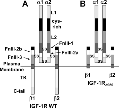
Figure 6.
IGF-IR structure. A and B, Diagram of the full-length IGF-IR (A) and C-terminally truncated IGF-IRΔ950 mutant (B). IGF-IR is a disulfide-linked heterotetramer consisting of an α2β2 assemblage in which the α-chain is entirely extracellular and the β-chain is a transmembrane protein that harbors a tyrosine kinase in its intracellular domain. L, leucine-rich repeat domain; Cys-rich, cysteine-rich; FnIII, fibronectin III; TK, tyrosine kinase domain.

