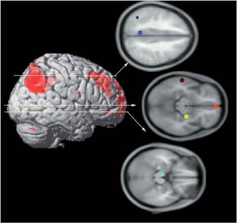
Fig. 2.
Six regions were included in the anatomical model; these regions were selected based upon prior studies of autobiographical memory retrieval (e.g. Maguire et al., 2001; Sharot et al., 2007). Regions included medial parietal cortex (encroaching upon posterior cingulated cortex and precuneus; center of ROI = −6, −50, 37; in blue), left temporoparietal junction (center of ROI = −40, −56, 43; in navy), medial PFC (center of ROI = 2, 55, −13; in orange), lateral middle temporal lobe (center of ROI = −60, −22, −10; in brown), right amygdala-hippocampal complex (center of ROI = 26, −12, −11; in yellow), and left hippocampus (center of ROI = −14, −18, −18; in turquoise).

