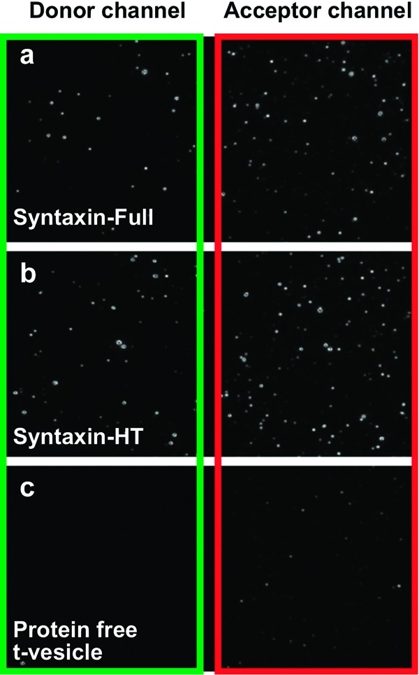
Figure 2.

Laser-excited (532 nm) images of single-vesicle fusion experiments with Munc18-1. Acceptor-labeled v-vesicles are directly tethered to the surface via biotin−neutravidin linker and the donor-labeled t-vesicles are added. Because the laser excites the acceptor only very weakly, bright fluorescent spots are seen only when the t-vesicles are present: (a) t-vesicles containing syntaxin-full and SNAP-25, (b) t-vesicles containing syntaxin-HT and SNAP-25, and (c) protein-free t-vesicles. Green and red rectangles denote the donor and acceptor emission detection channels, respectively. Panels a and b show docked t-SNARE vesicles in the donor channel and bright v-SNARE vesicles through FRET in the acceptor channel. Strong FRET signal demonstrates that binding of t-SNARE vesicles to the surface is specially achieved via interaction with the surface-immobilized v-SNARE vesicles. Panel c only shows dim v-SNARE vesicles in the acceptor channel without docking of t-vesicles, demonstrating that the nonspecific adhesion of the t-SNARE vesicles to the surface is minimal. In all experiments, 1 μM Munc18-1 was used.
