Figure 4.
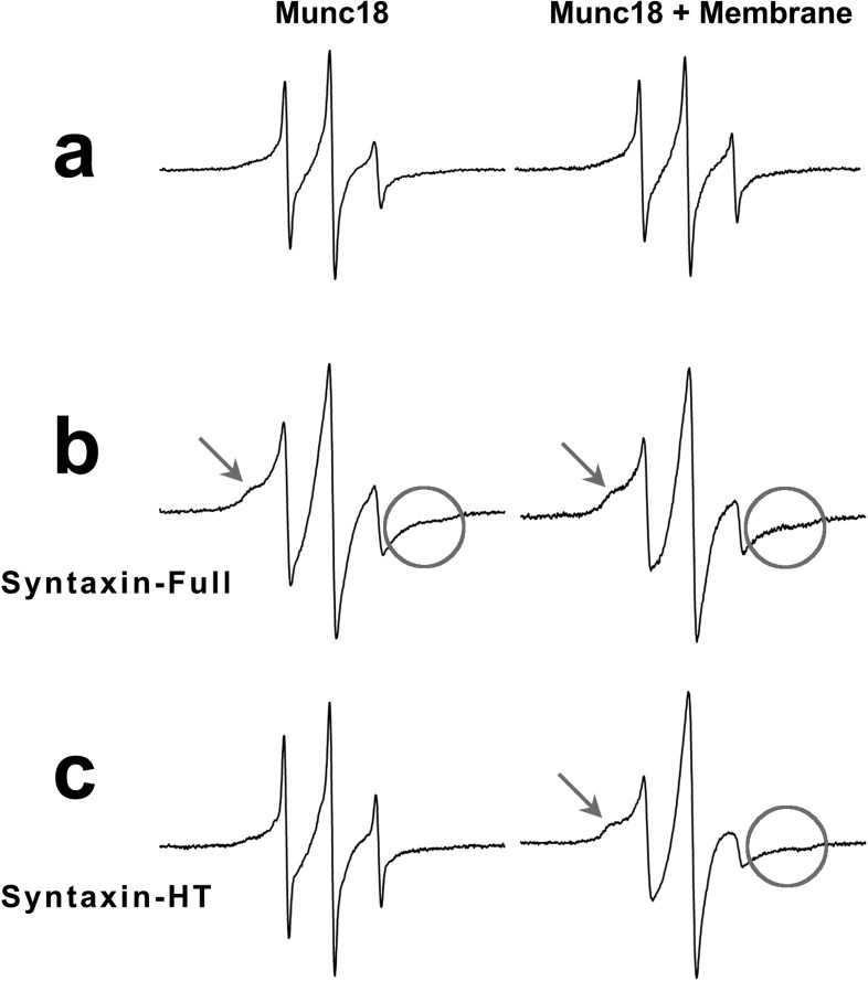
Electron paramagnetic resonance (EPR) spectra of Munc18-1. (a) Munc18-1 only (left panel) and Munc18-1with protein-free membrane in the fusion buffer (right panel). The same EPR sharp spectra indicate that there is no measurable interaction between Munc18-1 and membrane. (b) Munc18-1 with syntaxin-full protein (left panel) and Munc18-1with membrane-associated SNARE complex containing syxtaxin-full (right panel). Both EPR spectra are broadened in comparison to the spectrum of Munc18-1 only (a, left panel) indicating interactions. (c) Munc18-1 with syntaxin-HT protein (left panel) and Munc18-1 with membrane-associated SNARE complex containing syntaxin-HT (right panel). The sharp EPR spectrum on the left indicates that there is no interaction between Munc18-1 and syntaxin-HT, while the broadened EPR spectrum on the right suggests interactions. Arrows indicate the quaternary interaction between Munc18 and SNAREs, and circles show the spin−spin interaction due to the clustering of spin-labeled Munc18.

