Fig. 7.
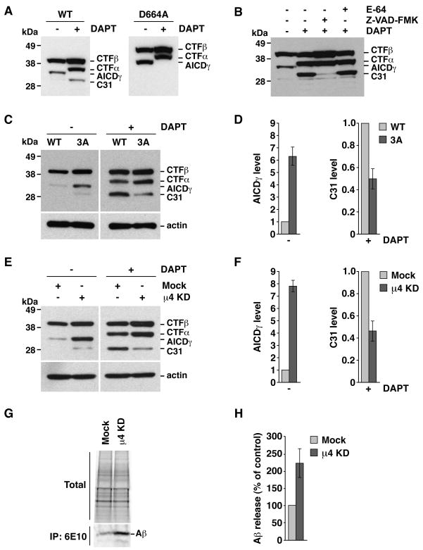
Altered processing of CTFβ upon disruption of the YKFFE-μ4 interaction. Experiments were performed as in Fig. 6, with the variations indicated below. The positions of molecular mass markers and different APP species are indicated. (A) Cells expressing normal CTFβ-GFP (WT) or CTFβ-GFP carrying a D664A mutation. (B) Cells expressing normal CTFβ-GFP were treated with 100 μM E-64, 200 μM Z-VAD-FMK and / or 250 nM DAPT. (C) Cells expressing normal CTFβ-GFP (WT) or CTFβ-GFP with the triple mutation, F689A, F690A and E691A (3A). (D) Mean ± SD of AICDγ and C31 levels from three experiments such as that shown in C. (E) Mock- or μ4-depleted cells (μ4 KD) expressing CTFβ-GFP. (F) Mean ± SD of AICDγ and C31 levels from three experiments such as that shown in E. (G) Mock- or μ4-depleted cells expressing normal CTFβ-GFP were labeled for 4 h at 37°C with [35S]methionine-cysteine, and the culture medium was subjected to immunoprecipitation with antibody 6E10 to Aβ. Proteins were analyzed by electrophoresis on Tricine 10–20% acrylamide gradient gels and fluorography. An aliquot of the culture media (total) is shown as loading control. (H) Mean ± SD from four experiments like that in G. Control: mock.

