
Figure 3.
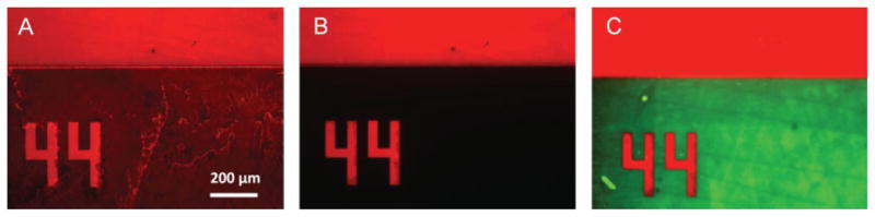
Fluorescent images of protein copatterning on PDMS using parylene. A: Initially, a Texas Red-BSA protein solution is incubated on the parylene membrane for 15–30 min. The protein (red) adsorbs both to the parylene and the exposed regions of the PDMS. B: After the parylene is peeled off, the adsorbed protein pattern remains on the substrate (red), while the unexposed regions are free of protein (black). C: A second protein solution of FITC-BSA (green) is incubated on the first pattern, selectively binding to the protein-free regions and creating a protein copattern. [Color figure can be viewed in the online issue, which is available at www.interscience.wiley.com.]